Purpose of the visit: why did you apply for a job shadowing mobility program within BNMI?
I had recently started a position in a microscopy core facility in Trondheim and was eager to expand my experience with advanced light microscopy methods, hence becoming able to offer more advanced support to our users. When I was approached by a new group of users that were interested in using our systems for FLIM and FRET experiments (for which my experience was limited), I thought the BNMI job shadowing program would be the perfect place to start. The aim of the visit was to gain hands-on experience with new imaging techniques, and at the same time to learn about facility management at other institutions, and to establish new connections in the Nordic countries.
Description of the work: what training did you get?
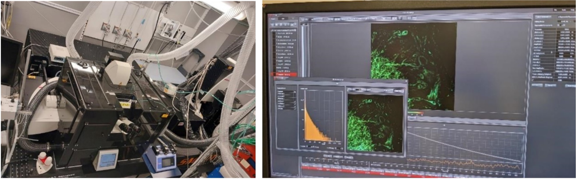
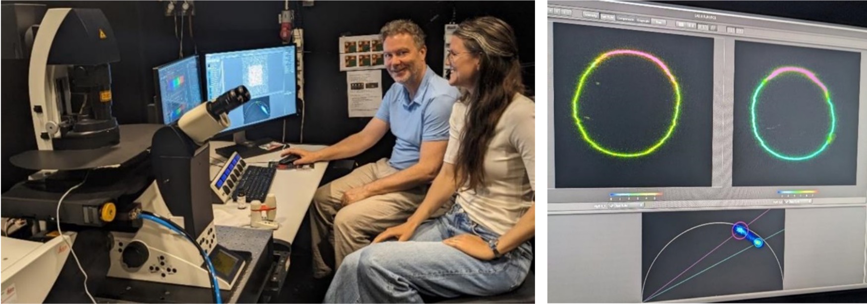
I visited the microscopy facilities ALM/SciLifelab and IVMSU in Stockholm, which are led by Hans Blom and Christiane Peuckert, respectively. At IVMSU, I had my first experience with a microscopy facility operating within the enclosed environment within an animal facility. Christiane Peuckert nicely demonstrated a FRET calibration workflow by mixing solutions of FITC and Alexa 555, and furthermore demonstrated FLIM recording and analysis on an octopus sample from the Norwegian fjords (Fig. 1). We also used the two-photon setup to perform label-free imaging of dinosaur bones, which emphasized the variety of samples and labelling (or absence thereof) one can meet at IVMSU. Finally, I got a very interesting demonstration of a surgical window implantation for intravital microscopy in mice.
At ALM/SciLifeLab, Hans Blom introduced me to STED, gated STED and Tau-STED (lifetime-filtered STED), which was an inspiration for us to advance super resolution support in our own facility (Fig. 2). Franziska Ragaller further performed live FLIM and phasor plot analysis in phase separating GUVs (the “soap bubbles”), while Francesca Pennacchietti introduced the characterization of photoconverting molecules with an accompanying shift in fluorophore lifetime. All the demonstrations were hands-on on the microscope, and I learnt a lot during our time together.
Outcome: did you acquire some new knowledge and skills?
ALM/SciLifeLab and IVMSU together cover a broad range of advanced methods in light microscopy. Hans Blom and Christiane Peuckert gave me a complete tour of their facilities, including lab spaces, analysis workstations and microscope instruments. At SciLifeLab I was also introduced to facility head Marta Carroni who showed me around the cryo-EM facility, much to the enjoyment of an electron microscopy enthusiast. Of special interest was how the different imaging facilities were physically organized, how different projects were evaluated before starting, and also how the different facilities emphasized different aspects of a microscopy workflow depending on their scope and user base.
All the practical training described above was performed with a high level of detail and an openness to questions that would make it possible for me to bring the practical experience back to the lab in Trondheim to support our users in FLIM, FRET and STED experiments. Furthermore, both of my hosts were happy to keep in touch in the future for possible collaboration projects or future support.
Feedback to other scientists on using BNMI’s available mobility support:
I would highly recommend other scientists to apply for BNMI’s job shadowing program. This unique opportunity allows microscopy facility staff to expand their expertise and build valuable connections across the Nordic countries. I believe this initiative can enhance the quality of microscopy facilities by strengthening our collective knowledge base, hence strengthening our contribution to the scientific community. On the personal side, it’s a lot of fun to learn new microscopy methods and to meet fellow microscopy enthusiasts!