1. Why did you apply for short term scientific mission support within BNMI?
Confocal microscopy is available in Iceland but currently there is no fluorescence microscope capable of super-resolution (SR). Although the confocal is a flexible and highly useful system, the absence of any locally available SR method limits what can be explored and studied with fluorescence microscopy for those who would be interested, as well as limiting the opportunities for knowledge transfer concerning these methods. BNMI’s objective of strengthening microscopy networks and collaboration in the Nordics, for instance through their short-term scientific mission (STSM) program, gave me the opportunity to use these otherwise unavailable methods to further explore our research questions, as well as providing funding for the exchange and our project.
2. What was your scientific project about?
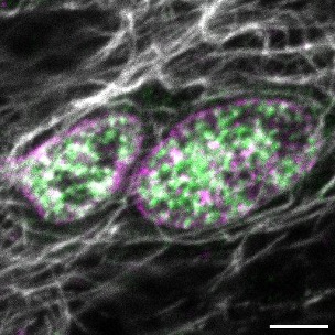
For the STSM we were primarily interested in observing our proteins of interest (POI) and microtubules in the neuromuscular junction (NMJ) of live Drosophila melanogaster larvae. Of equal importance was to explore the applicability of a STED microscope on our current sample preps and, if successful, image the consequence of POI knock-down on selected NMJ proteins.
3. What kind of support did you get?
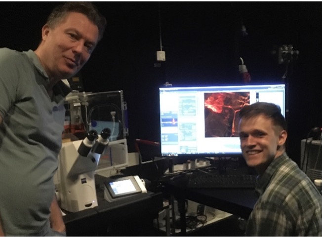
During the STSM I had a lot of support from the Advanced Light Microscopy (ALM) facility in Stockholm with using and training on their high-end microscopes and applying image deconvolution with their desktop licences. I also had a lot of support from academic collaborators at the Royal Institute of Technology (KTH) through the sharing of lab space and reagents. Finally, I was also able to house our banana flies with academic collaborators at Stockholm University (SU). All of this made it possible to do the needed work and I am very grateful for the excellent support I was shown and given during the STSM.
Scale bar 1 mm.
4. Did you get any new results?
The STSM was good experience and time well spent. We were able to ascertain the usefulness of different methods for our research questions and get meaningful super-resolved fluorescence imaging data with 3-color STED imaging.
5. What is your feedback for getting more scientists to use the available Bridging Nordic Microscopy Infrastructure STSM support?
I strongly encourage scientists in the Nordic countries to take advantage of the short-term scientific mission (STSM) program. Not only does it provide an opportunity to collaborate with other scientists within the Nordics, but it also allows for access to advanced microscopy infrastructure in the region. Additionally, the BNMI provides funding for short-term mission, which helped to greatly offset the costs of the journey. From my own personal experience, I can attest to the benefits and positive impact that an STSM can have on one’s research and career. Don’t miss this opportunity to advance your work and strengthen new and fruitful Nordic cooperations.
For further information about our mobility programs and funding opportunities like the STSM program, please go here.