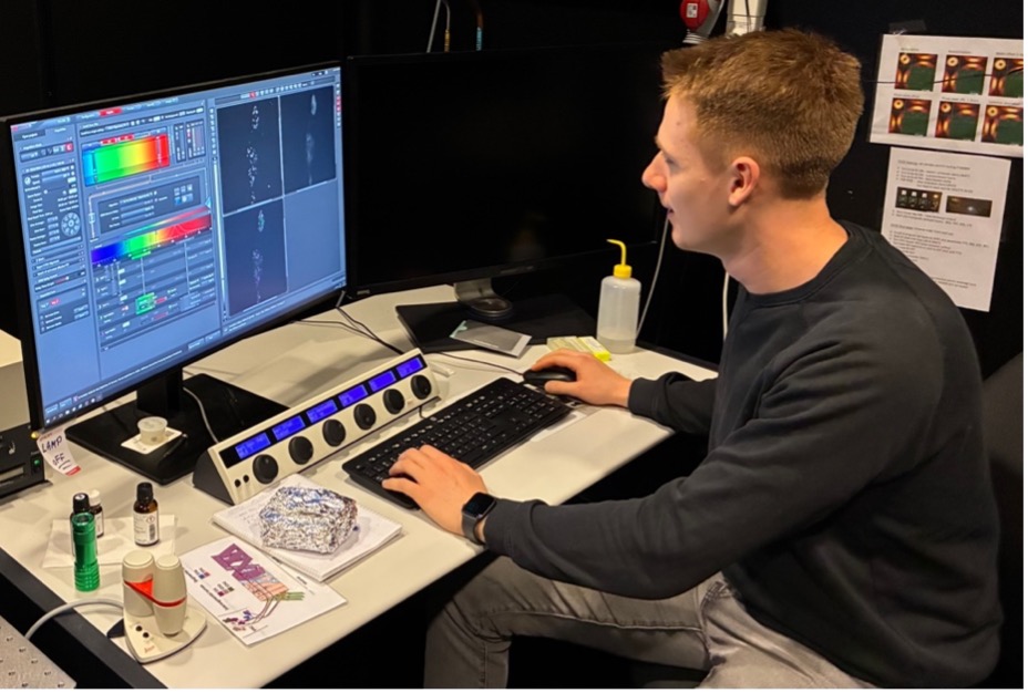
BNMI continues to support Nordic scientists with advanced microscopy and recently Gustav Schwenk (photo below), from the University of Iceland and member of Sigríður Rut Franzdóttir’s group, applied STED microscopy to further dissect synaptic proteins in fly larvae at the ALM facility in Stockholm.
How did you benefit from STED collaborative imaging support?
With the close support of Hans Blom and access to their STED technology (https://www.scilifelab.se/units/integrated-microscopy-technologies/), I was able to acquire images with high resolution and magnification that would not have been possible at my home institution. This enabled me to precisely analyze active zone structures in neuromuscular junctions of Drosophila larvae.
What do you think about getting support with imaging on a Nordic level?
I believe this is an excellent initiative that greatly benefits researchers who do not have access to such specialized equipment, as in my case. The combination of advanced imaging facilities and the expertise of highly competent personnel makes this support particularly valuable.
Would you change something on how BNMI can support projects?
No changes are necessary in my view. The current support options, including job-shadowing and short-term scientific missions provide sufficient assistance for researchers.