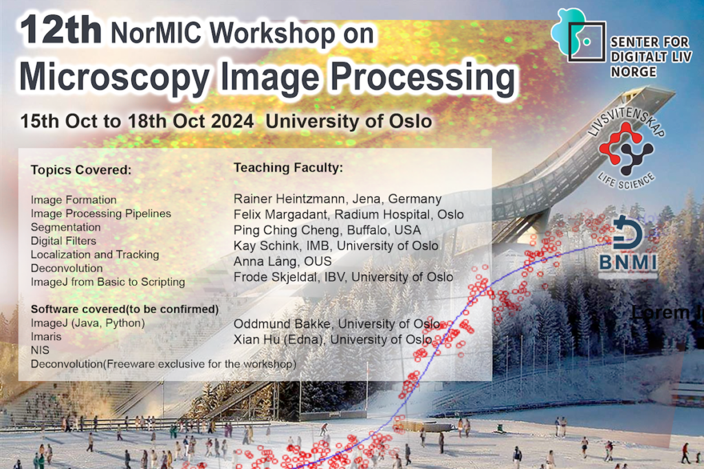
The NorMIC series of workshops aims to teach biological researchers (PhD students, engineers, postdocs and young PIs) the principles of biological microscopes and image processing.
Time and place: Oct. 15, 2024 – Oct. 18, 2024, Kristine Bonnevies Hus
Theory and Science in Biological Optical Imaging
We are placing special emphasis on scientific theory and concepts, rather than solely instructing on how to use specific microscopes or image processing software. We hope that this approach will enable the attendees to quickly develop an understanding of the framework of modern bioimaging sciences and improve their ability to navigate their specific bioimaging applications in the future. Additionally, we aspire to establish a local imaging community through these workshops.
Highlight of this workshop:
- As we have been organizing the BNMI symposium this spring, we did not hold the image acquisition NorMIC workshop. This upcoming workshop will be a hybrid, combining elements of both image acquisition and image processing. This means we will cover both topics in image acquisition and image processing in this workshop.
- The NorMIC Imaging Platform has recently acquired a state-of-the-art FLIM-STED microscope (Abberior, Facility Line), which is one of the most advanced STED microscopes in Scandinavian countries. During this workshop, we will spend time teach the optical principles and applications of FLIM and STED.
- In the near future, the NorMIC Imaging Platform will join the new Life Science Building‘s Shared Microscopy Facility. We are in the process of acquiring new imaging modalities, such as a Light Sheet Microscope and a High Content Screening Microscope. We will also dedicate time to cover these topics to prepare the local research community for the new systems that will soon be available.
The theory part of this course will cover: Super Resolutions Microscope Technology(SIM, Rescan, FLIM, STED, Array Detector), High Content Screening Imaging, CLEM , sample clearing, Image formation, processing workflow, segmentation, filters, de-convolution, rendering and visualization, classification.
As we also need to plan time to cover image acquisition topics in this course, the hands-on part will focus on teaching ImageJ, scripting ImageJ Macros, and performing deconvolution using software from the Heintzmann Lab. Both tools are freeware and open for academic use.
Registration
Tenative Teaching staff:
- Rainer Heintzmann (Leibniz Institute of Photonic Technology in Jena, Germany)
- Felix Margadant (UTMB, USA)
- Kay Schink (IMB, UiO)
- PC Cheng (State University of New York in Buffalo, USA)
- William Louch (HTH, University of Oslo, Norway)
- Joachim Mossige(RITMO, University of Oslo, Norway)
- Thomas Combriat (HTH, University of Oslo, Norway)
- Anna Lång (OUS, Norway)
- Frode Skjeldal (IBV, University of Oslo, Norway)
- Linda Haugen(IBV, University of Oslo, Norway)
- Oddmund Bakke (IBV, University of Oslo, Norway)
- Ingrid Kjos (Radium Hospital, Oslo University Hospital, Norway)
- Dominik Frei(Interinstrument AS)
- Edna, Xian Hu (Life Sciences Building Imaging Facility, UiO)
Organizing Committee: Edna, Xian Hu; Oddmund Bakke ; Felix Margadant ; Frode Skjeldal; Linda Haugen; Anna Lång
We receive financial support from: Center for Digital Life Norway , UiO:Life Sciences and Bridging Nordic Microscopy Infrastructure(BNMI)